Skip to content

Radiology and Mammography

In-House Radiology Services

Experience the Convenience and Comfort of Our In-House Radiology and Mammography Services At Atrium 

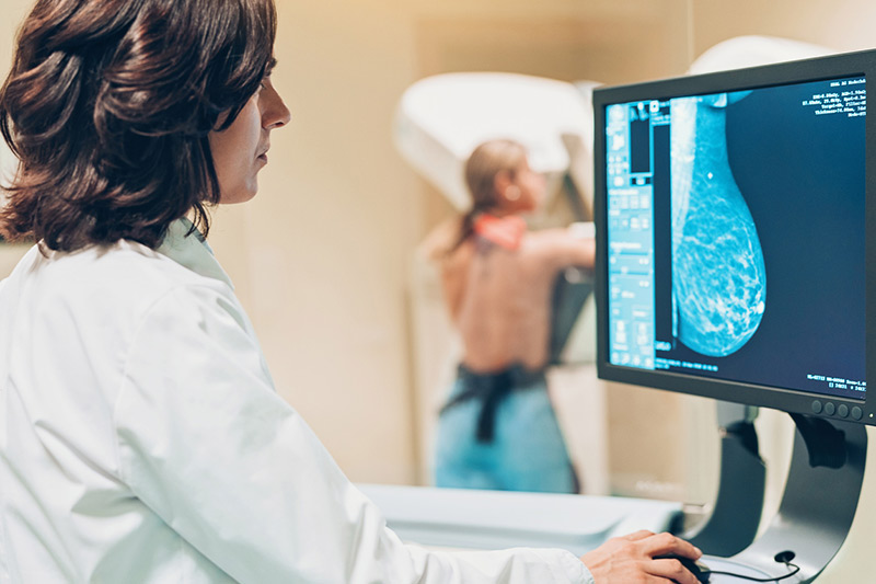
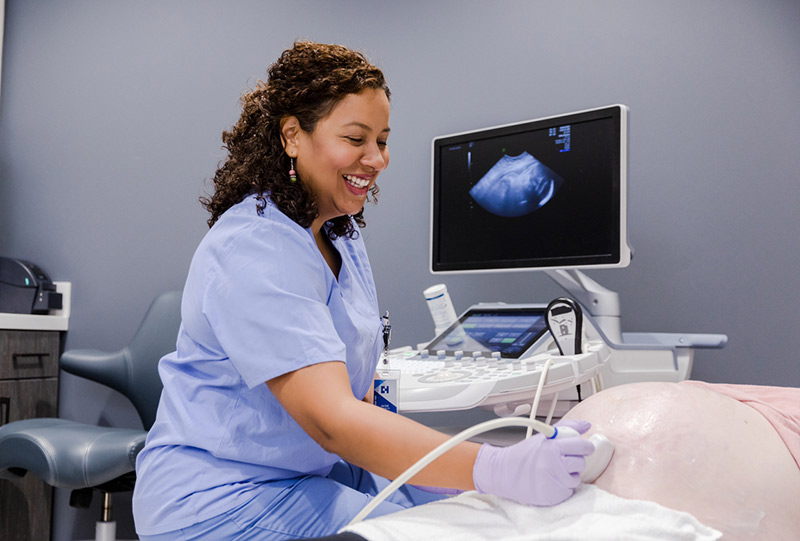
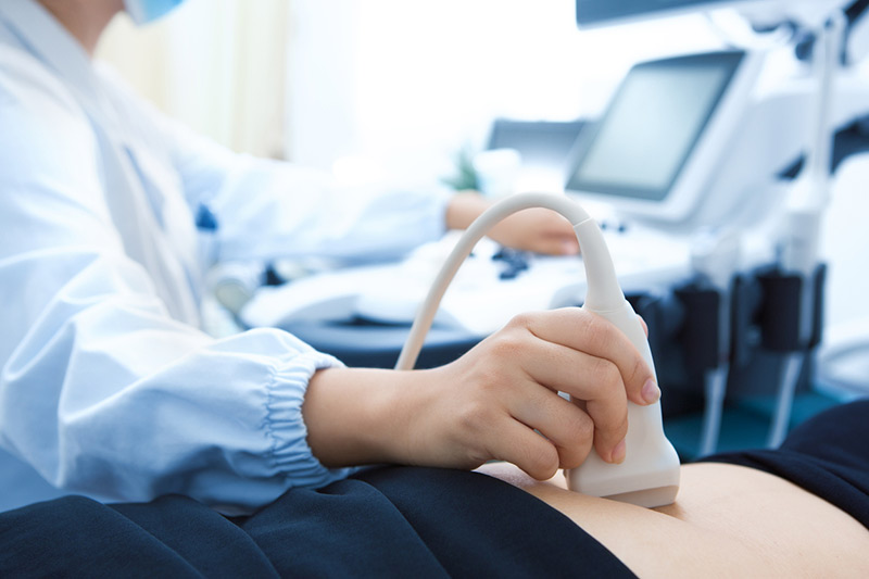
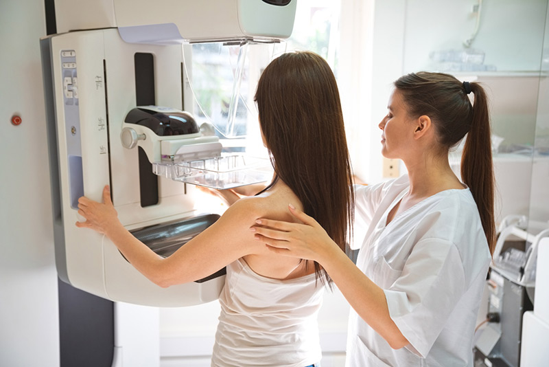
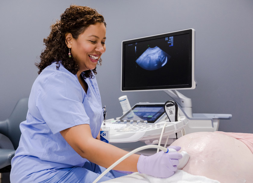
At Atrium, our advanced imaging technology includes fetal ultrasounds, diagnostic ultrasounds for breast, uterus, bladder and ovaries, as well as digital mammograms. We invite you to experience our pleasant surroundings and state-of-the-art equipment.

3D Mammography / Tomosynthesis Now Available

Atrium Ob/Gyn is pleased to offer our patients a breakthrough technology that revolutionizes how breast cancer is detected today - 3D Mammography, also known as breast tomosynthesis.

A 3D mammogram consists of multiple breast images taken in just seconds to produce a 3D image. The radiologist looks through the tissue one millimeter at a time seeing detail inside the breast in a way never before possible making breast abnormalities easier to see, even in dense tissue. It improves the radiologist’s ability to detect potential breast cancers by helping to pinpoint the size, shape and location of abnormalities and also enables the radiologist to distinguish harmless structures from tumors, leading to fewer false positives, fewer call-backs and less anxiety for women. Atrium Ob/Gyn is pleased to offer our patients a breakthrough technology that revolutionizes how breast cancer is detected today – 3D Mammography, also known as breast tomosynthesis.

The 3D mammogram is currently performed at the same time as the standard 2D digital mammography and does involve a minimal amount of additional radiation. The additional radiation is well within FDA’s recommended guideline limits for mammography. 

Helpful Mammography Videos

Atrium Patient Portal

Take advantage of the online services offered by our practice with the assurance that all of your information is encrypted and stored securely.

Mammography FAQs

The American Cancer Society and American Congress of Obstetricians and Gynecologists recommend that all women have a baseline screening mammogram between the ages of 35 and 40 and that beginning at the age of 40, women have an annual screening mammogram. Mammograms play a central part in the early detection of breast cancer because they can detect changes in the breast that may be early signs of cancer, but are too small or subtle to be felt.

A mammogram is an x-ray examination of the breasts, used to detect and diagnose breast diseases. Screening mammography is used as a preventive measure for women who have no symptoms of breast disease and usually involves two views of each breast. A diagnostic mammography involves additional views of the breast, and is used when an abnormality is found during a screening, or in women who have breast complaints, such as a breast mass, nipple discharge, breast pain, or skin irritation.

With digital mammography, the radiologist reviews electronic images of the breast, using high-resolution monitors. The physician can adjust the brightness, change contrast, and zoom in for close ups of specific areas of interest. Being able to manipulate images is one of the main benefits of digital technology. Another convenience is of digital mammography over film-based systems is it can greatly reduce the needs for retakes due to over or under exposure. This potentially saves additional time and reduces your exposure to x-rays. Digital mammograms produce images that appear on the technologist’s monitor in a matter of seconds. There is no waiting time for film to develop, which can mean a shorter time spent in the breast imaging suite.

There is no disagreement that early and accurate diagnosis of breast cancer is key to improving clinical outcomes. Mammography – and in particular, digital mammography – is now widely regarded as the gold standard for detecting early breast cancer disease. Mammography may detect cancer one and a half to four years before a cancer becomes clinically evident.

  • One in eight women living in the U.S will be diagnosed with breast cancer in their lifetime.
  • Breast cancer is the second leading cause of cancer death in U.S. women. It is the leading cause of death in 35 to 65 year old women.
  • Approximately 2.4 million American women with a history of breast cancer are alive today.

3D mammography combines a process known as “tomosynthesis” to produce a 3D mammogram in addition to a conventional 2D mammogram. Both image dimensions are important in screening for breast cancer. The 3D mammography technology allows the radiologist to see masses and distortions associated with cancers much more clearly in the 3D pictures. Calcification fields, which may be pre-cancerous indications, are more easily interpreted in the conventional 2D image. A good analogy for 3D mammography is like thinking of the pages in a book. If you look down at the cover you cannot see all the pages – but when you open it up, you can go through the entire book page-by-page to see everything between the covers. 3D mammography is designed with the same concept in mind.

Earlier Detection – 3D has been shown in clinical studies to be more accurate than conventional mammography alone in detecting cancers earlier. Better Visualization – 3D allows the radiologist to examine breast tissue layer by layer. Fine details are more clearly visible, no longer hidden by tissue above and below. Fewer Callbacks – 3D helps distinguish harmless abnormalities from real cancers, leading to fewer callbacks, fewer additional scans and biopsies, and less anxiety for patients.

The radiation exposure of 3D mammography is a little more than with 2D mammography. Yet the total dose of the 3D technique is still well within FDA safe limits and much less than the annual exposure everyone receives from natural sources, such as cosmic rays and the soil.

Our team of providers believe strongly that 3D technology is beneficial for all of our mammography patients – whether your breast tissue is fatty or dense, whether you have a history of breast cancer or not, and whether you are at high risk or not.

The 3D exam will feel very similar to a traditional digital mammogram. The equipment and compression of the breast are almost identical. What’s different is that the x-ray arm sweeps in a slight arc over the breast, taking multiple images in just seconds. High-powered computing will convert the images into a 3D image, which can then be examined by the radiologist one page or “slice” at a time.

AtriumGynecologyService

Gynecology

AtriumObstetricsService

Obstetrics

AtriumRadiologyService

Radiology

AtriumAdolescentCareService

Adolescent Care

How Much Do You Know About Breast Health?

When it comes to cancer prevention, knowledge is your best friend. When you get down to it, screening for breast cancer can increase your chances of finding the disease earlier. Whether you have implants or not, are nursing or not, or you’re pregnant or not: all women of appropriate screening age need to schedule their annual mammograms. We can help you get started

Remember To Schedule Your Annual Mammogram

Play Video